
Release time :2024-01-16
Source:support@yingchitech.com
Scan:1435
Clinical Support Department of Shenzhen Yingchi Technology Co.,Ltd.
In December 2023, Prof. Kai Yuan from Xidian University and Prof. Yifei Zhu from Hebei Medical University published a paper entitled “Electroencephalography microstates as novel functional biomarkers for insomnia disorder” in General Psychiatry (IF=11.9), the team collected daytime awake EEG data and Polysomnography (PSG) sleep data of patients with insomnia, which revealed that abnormal EEG microstates in patients with primary insomnia could be regulated by rTMS. The characteristics of the EEG microstate at baseline can also help to screen the people who are advantageous for rTMS intervention in insomnia.

Primary insomnia is not due to physical disease, mental illness, drugs, bad sleep environment or other factors caused by insomnia, is caused by genetic, environmental, psychological and behavioral factors together, so that the mind is in a state of excessive awakening results. Microstate analysis of the brain uses multi-channel EEG to collect EEG data, select variables of interest to define the instantaneous state of the system, and describe changes in brain activity according to changes in state characteristics (such as the duration or frequency of a specific state). Many neurological and psychiatric diseases exhibit time-dynamic changes of specific microstates, so microstate analysis is expected to help us to describe the hyperarousal state of insomnia and reveal a new electrophysiological mechanism of primary insomnia. At the same time, existing studies on rTMS have been proven to improve sleep quality in insomnia patients, but the relationship between sleep quality improvement and changes in EEG microstates is unclear, and whether individual differences in treated populations can be predicted by baseline microstate characteristics is rarely reported.
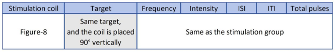
Two datasets were included in this study. The primary dataset included 40 healthy control patients and 60 patients with insomnia disorder. Insomnia patients were randomly divided into active stimulation treatment (n=30) and sham stimulation treatment (n=30). This dataset is used to explore abnormal brain microstates in patients with insomnia disorder and evaluate the impact of transcranial magnetic stimulation intervention on abnormal microstates in patients with insomnia disorder. The second dataset included 90 patients receiving true-stimulation therapy; It was used to verify whether the baseline microscopic state of patients with insomnia disorder could predict the outcome of 20-day active stimulation therapy. The inclusion and exclusion criteria for insomnia patients enrolled in the second dataset were consistent with those for the primary dataset. The stimulus parameters are as follows:

Polysomnography (PSG), Pittsburgh Sleep Quality Index (PSQI), Insomnia Severity Index (ISI), Epworth Sleepiness Scale (ESS), Beck Depression Inventory (BDI), Beck Anxiety Inventory (BAI), Mini-Mental State Examination (MMSE), Montreal Cognitive Assessment (MoCA)
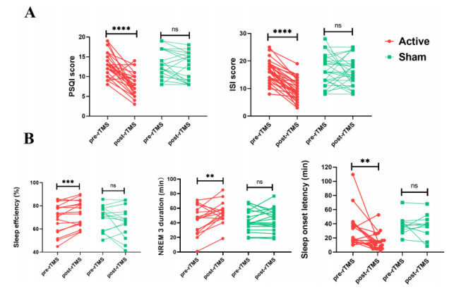
After treatment with TMS with active stimulation, patients with insomnia disorder showed significant improvement in subjective sleep quality and decreased PSQI and ISI scores (Figure A). The improvement in objective sleep quality was manifested by an increase in sleep efficiency (SE) and duration of non-rapid eye movement sleep stage 3 (NREM 3) and a reduction in sleep onset latency (SOL) (Figure B). In contrast, there was no significant change in subjective or objective sleep quality in the sham stimulation group. There were no significant differences in MMSE, MoCA, BDI and BAI scores in patients with insomnia disorder before and after active or shame TMS therapy.
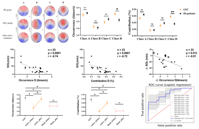
The results of microstate analysis showed that at baseline, the incidence and contribution of type D microstates in the insomnia disorder group were significantly lower than those in the healthy control group. The topography of microstates A and C in the insomnia disorder group was significantly different from that in the healthy control group. We found that the occurrence and contribution rate of type D microstates are negatively correlated with SOL. There are no significant differences in other microstate characteristics.
There were no significant differences in class C microstate topographic maps between insomnia patients and healthy controls after transcranial magnetic stimulation. The incidence and contribution of class D microstates in the insomnia patients group improved to levels close to those observed in the healthy controls. However, no such improvement was found in the spurious stimulation group. In the true stimulus group, the change of class D microstate was also significantly negatively correlated with the improvement of SOL.
The EEG microstate parameters of 86 insomnia patients were dimensionically reduced using PCA (EEG data of 4 patients were excluded due to quality issues), then a ten-fold cross-validation strategy was implemented to segment the dataset, which was then input into a logistic regression classifier for training and testing. The average prediction accuracy of the final baseline status characteristics was 80.13%.
The results showed that the incidence of class D microstate was lower in insomnia population, which was significantly correlated with sleep latency. In addition, some abnormalities in the microstate of the brain can be restored to normal levels by rTMS intervention, and the baseline microstate characteristics can predict the therapeutic effect of rTMS on insomnia. The EEG microstate can be used as a new electrophysiological indicator for insomnia, and it is expected to be helpful for screening the advantageous treatment population.
In the pathological process of insomnia disorder, the dynamic changes of brain activity play an important role. As a cheaper and more mobile medical device, EEG recording systems have been widely used to analyze dynamic changes in brain activity. Eeg microstate analysis has high time resolution and high retest reliability, and it can identify discontinuous and nonlinear changes of global brain functional state. The variation of microstate characteristic parameters and the relationship between microstate and brain functional network reflect many neurophysiological characteristics. The instantaneous global functional state of the brain is reflected in its electric field structure. The cluster analysis method consistently extracted four head surface brain electric field structures that were able to best explain the differences in spontaneous EEG recordings over time. These four structures, known as EEG microstates Class A, B, C, and D, are related to speech/speech, vision, subjective sensory-autonomic processing, and attention redirection, respectively. Our study found a reduced incidence and contribution of class D microstates in patients with insomnia disorder compared to healthy controls. This suggests that people with insomnia may not get enough rest at night, which can interfere with normal life during the day and impair attention and cognitive control. This study used PSG to objectively assess sleep quality in insomnia patients, which had not been used in previous EEG microstate studies of insomnia disorders. It extended this microstate analysis to reveal the pathology of insomnia disorders and found that EEG microstates can be used as biomarkers of sleep quality in insomnia patients, especially for SOL. In the future, cognitive assessment needs to be included for further verification.
In addition, the study has some limitations. First, in the study, patients with insomnia were not followed up, which made it impossible to assess the long-term effects of TMS on patients with insomnia. Secondly, the relationship between the higher order index of PSG or the coupling of different frequency bands of PSG and EEG microstates has not been fully discussed. Finally, clinical assessment was performed using the MMSE scale, which is not a comprehensive cognitive assessment. Future studies aim to address these limitations and further investigate the pathogenesis of insomnia disorders as well as the mechanism of action of rTMS to provide additional insights and improve treatment strategies for insomnia disorders.
Overall, the study is the first to examine the temporal dynamics and spatial topographic maps of four typical EEG microstates in patients with insomnia disorder following TMS treatment. PSG was also used to investigate the relationship between the microscopic state of insomnia patients and objective sleep indicators. In addition, baseline microstate time indicators, such as transitions between different microstates, help select potentially high responders to TMS therapy. These results provide new electrophysiological insights into the pathology of insomnia disorders and the possible mechanisms of rTMS for the treatment of insomnia disorders. The clinical significance of EEG in guided transcranial magnetic stimulation in the treatment of insomnia disorders is also emphasized.

1.This content is organized by the Clinical Support Department of Shenzhen Yingchi Technology Co.,Ltd. Criticisms and corrections are welcome. For reprint, please indicate the source.
2.Reference: Guo, Y., Zhao, X., Liu, X., Liu, J., Li, Y., Yue, L., Yuan, F., Zhu, Y., Sheng, X., Yu, D., & Yuan, K. (2023). Electroencephalography microstates as novel functional biomarkers for insomnia disorder. General psychiatry, 36(6), e101171. https://doi.org/10.1136/gpsych-2023-101171