
Release time :2022-12-30
Source:support@yingchitech.com
Scan:2318
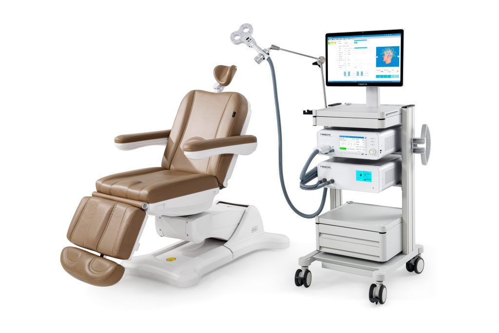
This is a blog post compiled and edited by the Clinical Support Department of Shenzhen Yingchi Technology Co.,Ltd. Wei Longxiao’s team at Tangdu Hospital of Air Force Military Medical University used M-100 ultimate, an rTMS device with a figure-of-eight stimulating coil (BY-90A) from YINGCHI, to conduct a research experiment on the mechanism of rTMS influence on the brain default function network of heroin addicts.
Heroin addiction is defined as compulsive heroin seeking and consumption behavior, despite awareness of severe negative consequences, which can lead to serious social and personal problems such as personal morbidity, economic loss, public health crisis, and overinduced mortality (Kolodny et al., 2015). Repetitive transcranial magnetic stimulation targeting the left dorsolateral prefrontal cortex has been shown to reduce craving levels in heroin addicts, but the neural mechanism by which it reduces craving levels is not well understood.
Menon (Menon, 2011) proposed a triple brain network model consisting of default mode network (DMN), salience network (SN), and executive control network (ECN), which is persistently abnormal in heroin addiction (Volkowet aL, 2011 ). The DMN mainly include s the medial prefrontal cortex (MPFC), posterior cingulate cortex (PCC), and bilateral inferior parietal lobule (IPL), involved in attention, self-monitoring, and introspective thinking (Greicius etal, 2003). The ECN includes the bilateral dorsolateral prefrontal cortex (DLPFC) and bilateral posterior parietal cortex (PPC), and is involved in attentional processing, working memory, and decision-making (Li et al, 2018).
Heroin dependent (HD) individuals exhibit abnormal brain activity and functional connectivity within and between the DMN and ECN. Longxiao Wei's team at Tangdu Hospital of Air Force Military Medical University performed functional magnetic resonance imaging (fMRI) to analyze the functional connectivity within and between the DMN and other brain networks in HD patients before and after 7 sessions of transcranial magnetic stimulation.
A total of 30 heroin addicts were included in the study, and data on craving and withdrawal symptoms as well as resting-state fMRI data were collected before and after the intervention. Meanwhile, 30 demographically matched healthy subjects who did not receive repetitive transcranial magnetic stimulation were included as a control group. This study focuses on the functional connectivity changes of the medial prefrontal cortex, posterior cingulate gyrus and bilateral inferior parietal lobule in the default function network core brain area of heroin addicts before and after the intervention. The authors defined four regions of interest (ROI) in the DMN, including the MPFC, PCC, and bilateral IPL. The authors conducted a paired t-test for HD (before treatment) vs HD (after treatment); two-sample t-tests for HD (before treatment) vs HC and HD (after treatment) vs HC.
All HD patients received 7 repetitive transcranial magnetic stimulation interventions at 10 Hz in one week. The rTMS group received 10Hz stimulation of the left DLPFC for 10min (5s stimulation time, 10s stimulation interval, 2000 pulses). An M-100 ultimate (YINGCHI) rTMS device with a figure-eight stimulating coil (BY-90A) was used.
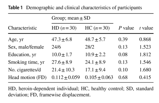
There were no significant differences between the HD (heroin dependent individuals) group and the HC (healthy controls) group in terms of age, years of education, duration of smoking, daily smoking or not, and head movement.
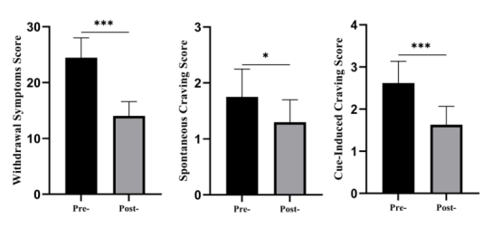
The above figure shows that the paired t test results show that after 7 courses of transcranial magnetic stimulation, the withdrawal symptom scores (t=4.31; P<0.001), spontaneous craving scores (t=2.27; P<0.05), and heroin-induced craving scores (t=4.36; P<0.001) were significantly decreased in HD patients.
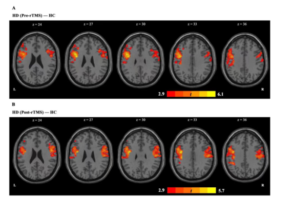
As shown in the figure above, in the PCC-based analysis, we found an increase in PCC-left inferior frontal gyrus and PCC-right anterior central gyrus coupling in the HD group compared to the HC group.
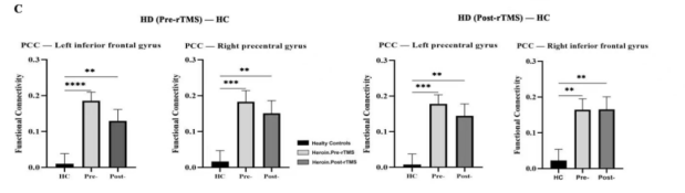
After the completion of a seven-course rTMS course, the increase in PCC-left inferior frontal gyrus coupling abnormalities was followed by a decrease in the HD group, and a similar trend was observed in the HC group. But after treatment, (HDvsHC) PCC-right precentral gyrus coupling persisted.
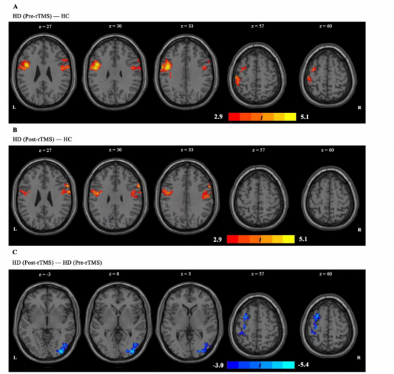
The upper panel shows the functional connectivity between the left IPL and the whole-brain network after transcranial magnetic stimulation, using two-sample t-test. Figure A shows that the left IPL-left precentral gyrus coupling and the left IPL-right inferior frontal gyrus coupling increased in the HD (before treatment) group compared with the HC group. Figure B shows that after rTMS treatment, the abnormal increase of left IPL-left precentral gyrus coupling and left IPL-right inferior frontal gyrus coupling in HD group was significantly reduced, and after 7 courses of rTMS treatment, the trend was close to that of HC group. Figure C shows that rTMS has a significant effect on left IPL-left middle frontal gyrus coupling and left IPL-right suboccipital gyrus coupling. In both regions, functional connectivity decreased significantly after 7 sessions of TMS.
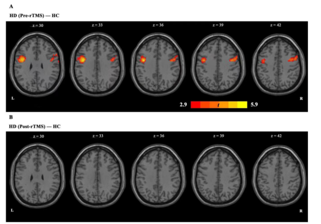
The upper image shows the functional connectivity between the right IPL and the whole-brain network after transcranial magnetic stimulation. Panel A shows an increase in right IPL-left precentral gyrus coupling and right IPL-right precentral gyrus coupling in the HD group (before treatment). Panel B shows that these abnormally increased connections were reduced after 7 sessions of TMS.
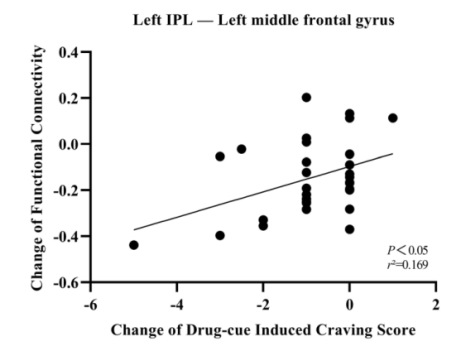
Regression analysis revealed a positive correlation between the change in functional connectivity between the left inferior parietal lobule IPL and the left middle frontal gyrus and the change in drug cue-induced craving.
Repetitive transcranial magnetic stimulation can modulate the functional connectivity between the default function network and the executive control network, and changes in the functional connectivity between the left inferior parietal lobule and the left middle frontal gyrus may play an important role in reducing drug cue-induced craving.
1.This content is organized by the Clinical Support Department of Shenzhen Yingchi Technology Co.,Ltd. Criticisms and corrections are welcome. For reprint, please indicate the source.
2.Reference: Jin, Long., Yuan, Menghui., Zhang, Wei., Wang, Lei., Chen, Jiajie.. Default mode network mechanisms of repeated transcranial magnetic stimulation in heroin addiction. Brain imaging and behavior, 2022.