
Release time :2023-03-27
Source:support@yingchitech.com
Scan:2164
Using transcranial magnetic stimulation (TMS) ----one kind of brain stimulation device,to regulate the function of brain neural circuits is becoming a new method for studying and treating various diseases. Previously, we have introduced how to improve the therapeutic effect of TMS from the perspective of temporal accuracy and situational accuracy. Today, we mainly share with you ways to improve the therapeutic effect of TMS in terms of spatial precision.
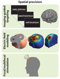
Figure 1: Spatial precision
Different diseases require different brain regions to be stimulated, and different stimulation methods are required. To improve the therapeutic effect of TMS in terms of spatial precision, it is mainly necessary to accurately place the coil on the corresponding stimulation target. A large number of relevant practitioners have continuously summarized various positioning methods during the use of transcranial magnetic therapy for diseases, ranging from rough to accurate, to cumbersome to intelligent. So, how exactly can transcranial magnetic pulses be accurately placed in the target brain region? Today, I will discuss with you the commonly used localization methods and procedures for transcranial magnetic stimulation.
Taking the target site of transcranial magnetic stimulation as the primary motor cortex as an example, there are currently various coil positioning methods available internationally, which can be roughly divided into the following six types [1], as shown in Figure 2 below:
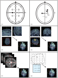
Figure 2: Six coil positioning methods
As shown in Figure 2 (1), the EEG 10-20 system is a standard electrode placement method developed by the International Brain Mapping Society. Its characteristics are that the electrode arrangement is proportional to the size and shape of the skull, and the electrodes are appropriately distributed at the main parts of the skull in standard positions. These electrode position markers reflect a relatively stable craniocerebral correspondence in a certain population.
Markers with the same name on different people's skulls can be considered to correspond to approximately the same anatomical region of the brain, such as the position of the C3 electrode, which corresponds to the left M1 region. When using a 10-20 brain electrode system to locate TMS coils in practice, it is often necessary to identify neural anatomical landmarks such as the nasal root point, occipital eminence, and bilateral anterior ear. This method is simple and convenient, and is currently widely used in clinical practice. However, the error between the roughly determined stimulation point and the actual effective site is often large.
As shown in Figure 2 (2), other brain regions with specific positional relationships are located based on functional brain regions that can be reasonably and conveniently located, such as M1. Taking the location of the dorsolateral prefrontal cortex as an example, in practice, the central location of M1 is determined by measuring motor evoked potential (MEP) using TMS, and then identified as the dorsolateral prefrontal cortex region 5 centimeters forward along the central axis. Similarly, similar empirical methods can be used to identify brain regions such as the premotor cortex and the sensory cortex.
This method is the standard method for determining M1, but it assumes that the relative position of each brain region is absolute, ignoring the significant individual differences in the anatomical position of the human brain.
As shown in Figure 2 (3). This type of neural navigation system collects images of the subject's head structure in advance, typically high-resolution T1 magnetic resonance images, and uses an external near infrared or optical navigation system to track the position of the stimulation coil.
Localization technology is achieved by registering magnetic resonance image information and computer vision images. Specifically, after using the MRI head scan image of the subject's three-dimensional head model, the subject's MRI structural image is used to reconstruct the three-dimensional model of the patient's head, and then an optical navigation system (such as Quiks Vision,Yingchi) is used to collect neuroanatomical landmarks of the head, track the position of the TMS stimulation coil in real-time, and the navigation system retrieves MRI imaging data for three-dimensional model reconstruction, After reconstruction, a doctor will designate the stimulation point based on professional knowledge and experience, guide the coil fixation position, and make the coil stimulation focus coincide with the designated stimulation point.
To achieve TMS positioning. This method is currently the most commonly used method in neuronavigation technology, and can well achieve accurate positioning of the anatomical position of the brain. However, it ignores the inconsistency between the anatomical position of the human brain and the functional position of the brain region, as well as the complexity of the projection position of the anatomical position on the scalp of the brain.
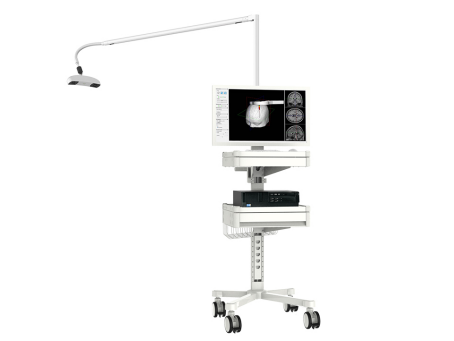
Figure 3: Yingchi TMS 3D Navigation System (Quiks Vision)
As shown in Figure 2 (4), generally based on the positioning method based on magnetic resonance structural images, the difference lies in not only scanning high-resolution T1 structural images of the subject, but also performing functional magnetic resonance imaging (FMRI), scanning the subject's functional magnetic resonance image, processing the functional magnetic resonance image, obtaining the activated brain regions for a specific brain functional stimulation task, and then determining the spatial coordinates of the stimulation site.
Specifically, taking the positioning of M1 as an example, first, a functional magnetic resonance experiment is designed according to the experimental paradigm, and functional magnetic resonance images of subjects during finger movement tasks are collected. Then, functional magnetic resonance images are processed to obtain statistical parameter maps, and the coordinates of the primary motor cortex are determined based on the activated brain regions.
Then, the coordinates are projected onto the constructed three-dimensional head model of the subject, and the stimulation points are determined. The navigation system guides the fixed position of the coil, so that the coil stimulation focus coincides with the designated stimulation points to achieve TMS positioning. The problem with this method is that the localization of brain function at the individual level may not be accurate.
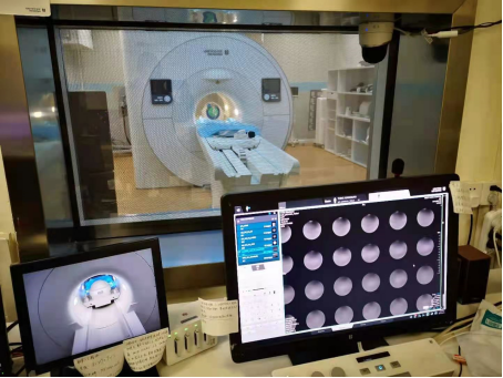
Figure 4: Real time image of Yingchi MRI coil in magnetic resonance water model experiment
As shown in Figure 2 (5). This method compensates for the disadvantage that individual functional magnetic resonance experiments cannot determine the functional regions of the brain. Specific TMS stimulation tasks were conducted with a group as the research object. First, select a specific population to perform a functional magnetic resonance experiment or positron emission tomography (PET) experiment under a specific stimulation task, and then search for a specific activated brain region based on the functional image of the population to determine the coordinates of the stimulation site.
In theory, this method combines individual magnetic resonance structural information and group functional magnetic resonance information, and can accurately determine the spatial coordinates of the stimulation site. However, in practical work, this method has problems such as high cost and difficulty in finding suitable homogeneous groups.
As shown in Figure 2 (6). This method does not perform functional magnetic resonance experiments, but requires the acquisition of T1 structural images of subjects. Through literature search for FMR/PET research results, the coordinates of specific populations and specific functional brain regions are determined through meta-analysis, and then directly applied to three-dimensional models reconstructed from subjects' magnetic resonance structural images and computer vision images. The coil positioning is guided through navigation systems.
The solution of the TMS localization problem relies on information from three levels of the subject's head: scalp shape, brain anatomical structure, and brain functional area localization [2]. Conventional TMS positioning and navigation systems either assume a corresponding relationship between the shape of the scalp and the anatomical structure of the brain, or assume a corresponding relationship between the anatomical structure of the brain and the functional regions of the brain.
These assumptions themselves have some truth. However, in the actual operation of TMS, the localization of stimulation coils should take into account the individual differences of the subjects and the limitations of the hypothesis itself. Therefore, relying solely on the anatomical knowledge possessed by doctors for direct positioning is a rough use of the subject's head shape information, assuming that there is a direct correspondence between the subject's scalp shape and brain functional areas.
If you use a positioning cap or EEG cap labeled with functional areas, it is also directly assumed that there is a direct correspondence between the scalp shape and the functional areas of the brain, ignoring the anatomical structure information of the brain. The error between the stimulation point roughly determined based on the subject's head shape and the actual effective stimulation site is often large, and the repeatability is poor [3].
A large number of researchers are exploring more possible methods in this direction to achieve the best therapeutic effect.
[1] Sparing R, Buelte D, Meister I G, et al. Transcranial magnetic stimulation and the challenge of coil placement: a comparison of conventional and stereotaxic neuronavigational strategies [J]. Human Brain Mapping, 2008, 29(1): 82-96.
[2] Weiduschat N, Habedank B, Lampe B, et al. Localizing Broca’s area for transcranial magnetic stimulation: comparison of surface distance measurements and stereotaxic positioning [J].Brain Stimulation, 2009, 2(2): 93-102.
[3] Hui Wang,A Study of Transcranial Magnetic Stimulation Coil Positioning.Journal of Integration Technology,2013,2(4).